
Cyanotic Heart Disease: Clinical Pathophysiology, Flow Classifications, and Management Protocols
Cyanotic heart disease encompasses severe structural congenital cardiac anomalies that disrupt normal blood oxygenation. This comprehensive medical review details the mechanical right-to-left shunts that deliver desaturated blood directly into systemic circulation. It systematically categorizes these lesions based on pulmonary and systemic blood flow dynamics. Furthermore, the text addresses life-sustaining patent ductus arteriosus dependencies, acute pharmacological interventions, definitive surgical corrective pathways, and long-term surveillance protocols for adult survivors.
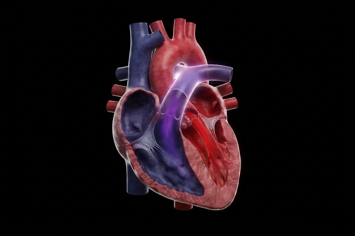
Table of Contents
-
Clinical Definition of Cyanotic Heart Disease
- Cyanotic heart disease defines a specific category of structural heart abnormalities present at birth. These malformations fundamentally disrupt the normal, sequential pathways of the human cardiovascular system.
- In a healthy heart, deoxygenated systemic venous blood returns directly to the right-sided chambers. The right ventricle pumps this unoxygenated blood into the pulmonary artery toward the lungs. The lungs perform gas exchange, fully oxygenating the blood before it returns left.
- Finally, the left ventricle forcefully pumps this oxygenated blood out into the systemic aorta. This efficient setup ensures that peripheral tissues receive a highly saturated, reliable supply of oxygen.
- In patients with cyanotic heart disease, this structured systemic-to-pulmonary pathway is deeply compromised. Specific structural anomalies cause poorly oxygenated blood to bypass the pulmonary tissue entirely and enter systemic circulation.
"Cyanotic congenital heart disease represents an anatomical failure of the in-series circulatory system, forcing deoxygenated blood into systemic pathways."
Consequently, the tissues and organs throughout the body receive blood with abnormally low oxygen saturation. This pervasive state of low arterial oxygen saturation clinically manifests as visible central cyanosis.
Cyanosis presents as a distinct, bluish or purplish coloration of the skin and core mucous membranes. It is detectable when absolute deoxygenated hemoglobin in peripheral capillaries exceeds roughly 3 to 5 g/dL.
-
Core Pathophysiology and Hemodynamic Mechanisms
The underlying pathophysiological hallmark of cyanotic heart disease is the development of a right-to-left shunt. This structural shunting
occurs when deoxygenated blood is diverted from the right heart directly to the left.
For a right-to-left shunt to occur, two specific anatomical conditions must be met concurrently:
- There must be a physical pathway or communication linking the right and left sides of the heart.
- The blood pressure within the right-sided chambers must exceed the pressure on the left side.
Alternatively, a severe physical obstruction must block the normal forward flow of blood into the lungs. When right-sided outflow tracts are heavily obstructed, the right ventricular pressure escalates dramatically to overcome resistance.
Blood then naturally takes the path of lowest resistance through any existing septal defects. This unoxygenated blood mixes directly into the left atrium or left ventricle, diluting the systemic oxygen pool.
The heart then ejects this highly desaturated, mixed blood into the aorta to supply the body. In other complex defects, a single common cardiac chamber receives both systemic and pulmonary venous blood returns.
This setup causes complete, obligatory mixing of oxygenated and deoxygenated blood streams within that single zone. A third major pathological mechanism involves the development of completely isolated, parallel circulatory loops.
In this parallel arrangement, oxygenated blood continuously recirculates through the lungs while unoxygenated blood recirculates through the body. This parallel arrangement prevents life-sustaining oxygen from reaching systemic organs without additional mixing points.
Reviewed by the LibraryMedicine.com Clinical Editorial Board. Our authors are practicing physicians and medical educators dedicated to delivering evidence-based, up-to-date clinical guides.








Comments (0)