
Inguinal Hernia: Classification, Clinical Features, and Surgical Management
An inguinal hernia occurs when abdominal contents protrude through the inguinal canal, representing 75% of all anterior abdominal wall hernias. This article provides a comprehensive overview of direct and indirect inguinal hernias, highlighting risk factors such as male gender and increased intra-abdominal pressure. It details key clinical features, including the classic groin lump and cough impulse, alongside diagnostic approaches and evidence-based management strategies. The content covers surgical interventions—open versus laparoscopic repair—and discusses complications like strangulation and obstruction, offering a complete clinical resource for medical professionals and students.
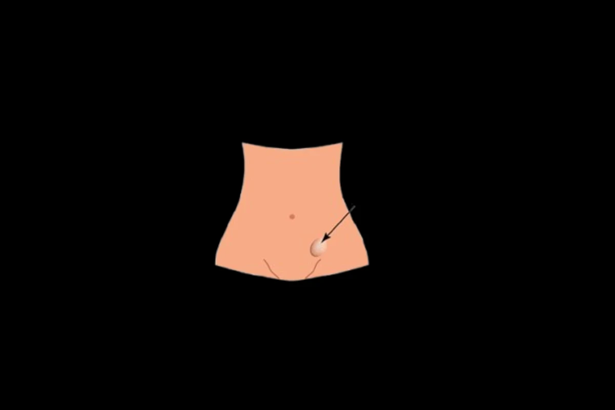
An inguinal hernia is defined as the protrusion of abdominal cavity contents into the inguinal canal. As the most common type of hernia, they account for approximately 75% of all anterior abdominal wall hernias, with a prevalence of 4% in individuals over 45 years of age. Understanding the anatomy, classification, and clinical management of inguinal hernias is essential for any clinician involved in general or emergency surgery.
Classification of Inguinal Hernias
A hernia represents the protrusion of an organ or tissue through the wall of the cavity that normally contains it. Inguinal hernias specifically involve abdominal contents passing into the inguinal canal, with potential extension into the scrotum. They are primarily divided into two subtypes:
-
Indirect Inguinal Hernia (80%): The bowel enters the inguinal canal via the deep inguinal ring. In younger patients, this results from incomplete closure of the processus vaginalis (a peritoneal outpouching that allows for testicular descent), making this subtype largely congenital in origin.
-
Direct Inguinal Hernia (20%): The bowel enters the inguinal canal directly through a weakness in the posterior wall of the canal, known as Hesselbach’s triangle. These occur more commonly in older patients, often secondary to abdominal wall laxity or chronically elevated intra-abdominal pressure.
The two types can only be reliably differentiated during surgery by identifying the inferior epigastric vessels: indirect hernias lie lateral to these vessels, while direct hernias lie medially. In pediatric cases, hernias are nearly always indirect.
Risk Factors
Several key factors increase the risk of developing an inguinal hernia:
-
Male gender
-
Increasing age
-
Elevated intra-abdominal pressure (from chronic cough, heavy lifting, or chronic constipation)
-
High body mass index (BMI)
Clinical Features
The most common presenting symptom is a lump in the groin. For reducible hernias, this lump initially disappears with minimal pressure or when the patient lies down. Mild to moderate discomfort may be present, often worsening with activity or standing.
If a hernia becomes incarcerated, the lump becomes painful and irreducible. Patients may present with features of bowel obstruction if the bowel lumen is blocked, or with signs of strangulation if the blood supply becomes compromised.
On examination, key features to note include:
-
Cough impulse: An irreducible hernia may not exhibit this.
-
Location: Inguinal hernias appear superomedial to the pubic tubercle (femoral hernias appear inferolateral).
-
Reducibility: On lying down or with gentle pressure.
-
Scrotal extension: Assess whether you can get above the mass and if it is separate from the testis.
Differentiating direct from indirect hernias on examination is often unreliable. Theoretically, reducing the hernia and applying pressure over the deep inguinal ring before asking the patient to cough can help—protrusion despite occlusion suggests a direct hernia, while no protrusion suggests an indirect hernia. However, definitive differentiation is only achieved intraoperatively.
Differential Diagnosis
Differential diagnoses for a groin lump include:
-
Femoral hernia
-
Saphena varix
-
Inguinal lymphadenopathy
-
Lipoma or groin abscess
-
If the mass extends into the scrotum: hydrocele, varicocele, or testicular malignancy
Investigations
Inguinal hernia is primarily a clinical diagnosis. Current Royal College of Surgeons guidelines recommend imaging only when diagnostic uncertainty exists or to exclude other pathology. Ultrasound is the recommended first-line imaging modality in the outpatient setting. For patients with features of obstruction or strangulation, CT imaging is required.
Management
Elective Management
Any patient with a symptomatic inguinal hernia (significant mass or discomfort) should be offered surgical intervention. The annual risk of strangulation for an inguinal hernia is approximately 2%. Importantly, one-third of patients with an inguinal hernia will remain asymptomatic, particularly those identified incidentally on imaging. In these cases, discussions should focus on the likelihood of future intervention and recognition of strangulation symptoms.

Dr. Emmanuel Hirwa BAKUNDA is a Medical doctor and technology innovator dedicated to transforming medical education through digital solutions. With a clinical background in General pediatrics and child health, Dr. Bakunda combines frontline patient experience with a passion for health informatics and e-learning platforms.













Comments (0)